What can X-ray software offer?
Today, software for digital X-rays, such as our proprietary EXAMION X-AQS X-ray software, must be able to offer all-round solutions. But what does this mean exactly? If you opt for image generation software such as EXAMION's X-AQS, it can be used to map the entire X-ray exposure workflow. The software manages every imaging parameter via the X-ray generator, controls the individual components of the X-ray system and optimises and analyses the radiological image data. In addition, the software saves the dose report and the X-ray log, and last but not least, it archives the X-ray images as well as the patient and order data.
Using X-AQS as an example, this specifically means that the software automatically and reliably manages the administrative load of most of the relevant steps in the entire X-ray process:
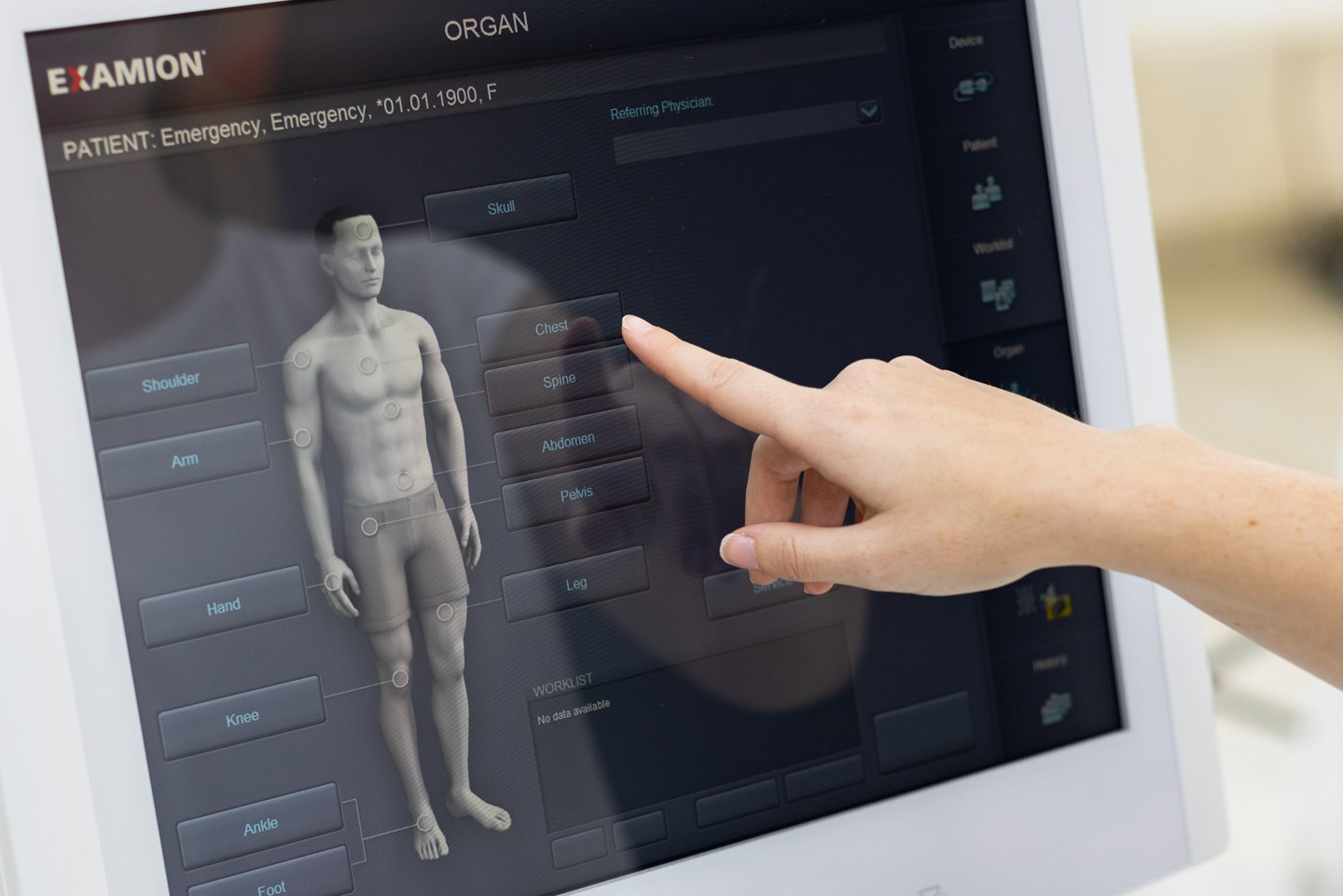
With the medical indication of an X-ray examination, patient data can be accessed directly via the X-AQS database and the user can select the desired organ display in the organ overview. The so-called software worklist of the shows all current X-ray orders so that you can access the respective patient entry to start the corresponding X-ray exposure.
The X-ray generator and all of the relevant parameters such as high voltage (kV), current (mA) and exposure time (ms) are managed according to the choice of organ.
Once these parameters are entered, the X-AQS provides visual support for correct patient positioning and summarises all the information and settings made so far on the exposure screen for a final check. Among other things, X-ray images of the patient that have already been taken can also be viewed at this point. After initiating the X-ray exposure, the software manages of individual components to ensure that the desired configuration is maintained.
The digital image preview, including post parameters, is then available to you immediately after the X-ray image has been acquired. All relevant data and values, including the corresponding dose report, as well as detector status and charge level are saved at this stage.
During automatic digital image post-processing, the X-ray software now takes on the critical role of optimising and analysing radiological image data. This ensures that X-ray images are output in a format that can be used for diagnostic purposes.
For more in depth analysis, you can also access extensive processing functions and diagnostic tools, such as gradation adjustments or special measurements, at all workstations.
The digital X-ray images are then archived via the PACS (picture archiving and communication system), which is also integrated within the X-AQS software. Images can also be transferred to a third-party PACS via a DICOM interface, and it is even possible to connect third-party devices. The externally generated image data can then be saved in the PACS.
