
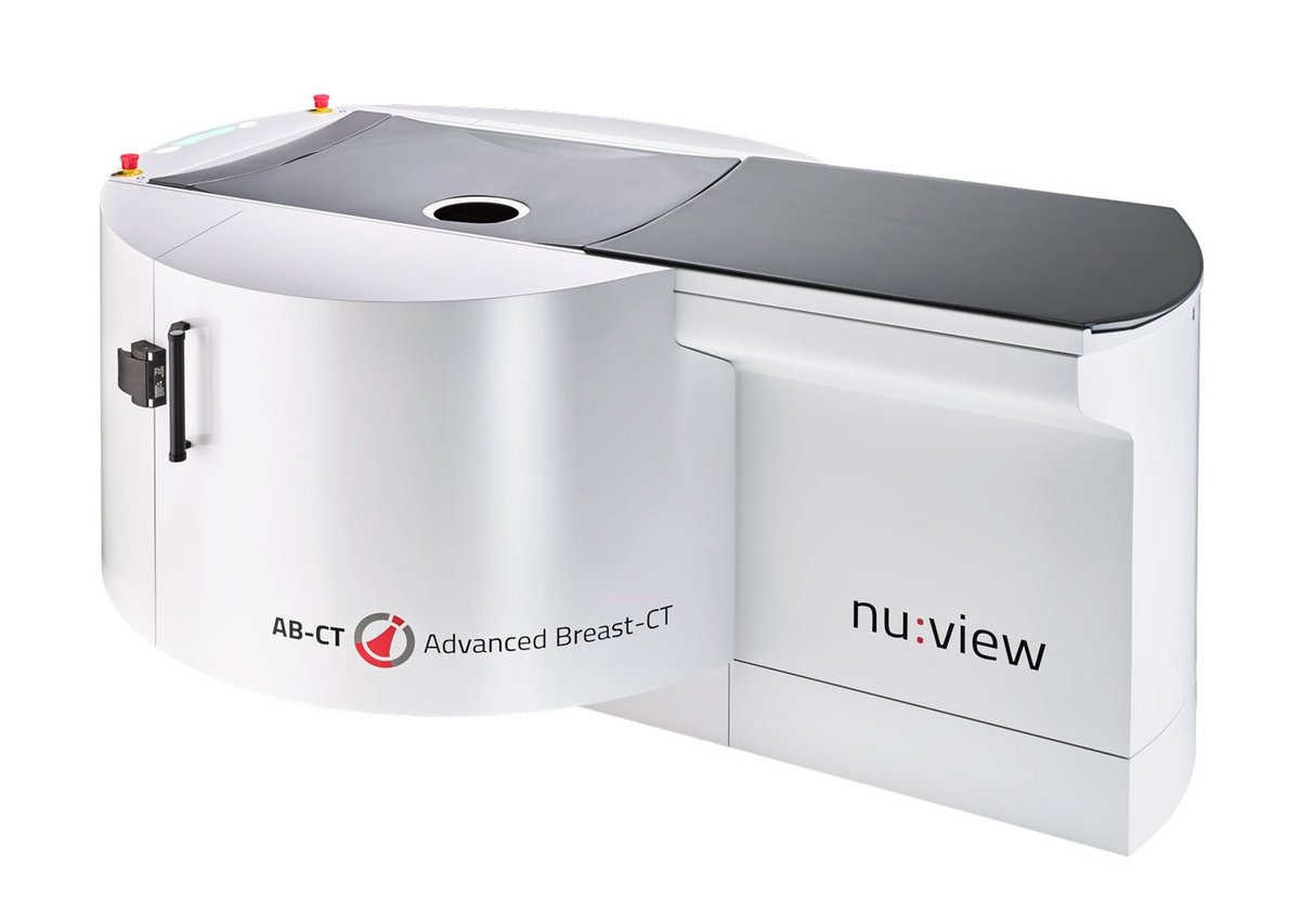
AB-CT nu:view
The nu:view from AB-CT is the world's first breast computed tomography (CT) scanner based on spiral CT technology and an imaging system that permits diagnosis of breast cancer in its very early stages. The system is characterised by excellent image resolution at low radiation doses with short scanning times.
In contrast to earlier detector technologies, nu:view converts each X-ray photon directly into an electrical pulse using cadmium telluride (CdTe) photon-counting detectors. nu:view is thus able to deliver the highest image quality with minimal dose requirements. For the first time, true 3D visualization of all parts of the female breast can now be acquired in a single image. The overlay-free images allow for excellent soft tissue differentiation. Up to 2000 projection images are created during a single 360° rotation around the female breast, and a complete scan takes only 7-12 seconds.
- High isotropic spatial resolution
- True 3D images of the breast without image overlays
- Excellent soft tissue differentiation
- Optimised for short scan times with an acquisition time of 7-12 seconds per scan
- Low patient dose during mammography scans
- Increased patient comfort – no compression of the breast is necessary
- CE marking, fully-integrated clinical information systems (RIS & PACS)


High patient comfort
nu:view breast CT offers a very high level of patient comfort, since the full examination is conducted without breast compression. For this purpose, the patient lies in the prone position (chest-down) on the examination table, the breast to be examined is placed comfortably over the aperture and no compression needs to be exerted.
There are no restrictions in terms of age, gender or other conditions of the breast for patients able to undergo examination.


X-ray tube
- Focal spot size 0.4 (IEC 60336)
- Tube voltage 60 kV
- Current 5-125 mA
- Power: up to 7.5 kW
- Filtering: 3 mm Al (equivalent)
Scan
- Spiral cone beam CT scan
- Up to 2000 projections in 360°
- Acquisition time 7-12 sec per scan
- Low patient dose for diagnostic mammograms
- No breast compression
Detector
- Type: photon counting detector (direct conversion)
- Sensor: CdTe, 0.75 mm thickness
- Pixel size: (0.1)mm²
- Detector area: approx. 280 x 50 mm²
- Frame rate: up to 1000 Hz
Reconstruction
- Fully isotropic high spatial resolution
- Measurement range: Ø 200 x 160 mm
- Voxel size: (0.15)mm³
- Reconstruction algorithm with filtered back projection
Find out more about nu:view in current publications
Published in Frauenheilkunde und Geburtshilfe 2026 (Language: English)
This experimental phantom study shows for the first time that the newly developed SBCT-compatible biopsy unit closes the previous diagnostic gap and enables both vacuum-assisted biopsy (VAB) and the marking of detected findings (especially microcalcifications and round foci) after precise three-dimensional target planning with the help of SBCT.
Published in Radiologie Magazin 4-2025 (language: German)
An interview with Prof. Thomas Frauenfelder, University Hospital Zurich, Dr. Karsten Ridder, MVZ Uhlenbrock, and Dr. Felix Althoff, AB-CT, on embedding the technology in clinical workflows, the importance of photon-counting technology, and the response of patients and referring physicians to breast CT.
published in Radiologie Magazin 1-2020 (Language: German)
In our news section you can read an article published in the Radiologie Magazin.
It reports in detail on the experiences of the University Hospital Zurich, where nu:view is used.
published in DIEurope (Feb/March 2020) (Language: English
A review article published in Diagnostic Imaging Europe (DIEurope) provides a comprehensive overview of nu:view.
The article clarifies questions such as "How is it possible to achieve high-resolution true 3D images at low dose?" and outlines the key benefits of the breast CT system for radiologists and patients. In addition, experience already gained with nu:view from clinical practice is shared.
published in DIEurope (Nov 2020) (Language: English)
Another article from Diagnostic Imaging Europe (DIEurope) introduces the radiology practice MVZ Prof. Dr. Uhlenbrock & Partner led by Dr. Karsten Ridder, the radiologist responsible for breast imaging. He talks in detail about the innovative nu:view breast CT system used in the practice and shares his experiences.
published in DIEurope (Jun 2021) (Language: English)
You can also learn more about the use of the breast CT system in patients with breast implants in Diagnostic Imaging Europe (DIEurope). Prof. Andreas Boss, senior physician in charge of breast imaging at the University Hospital Zurich, reports.
published im Radiologie Magazin 2-2022 (Language: German)
In the issue "Mammadiagnostik Special" of the Radiology Magazine 2-2022, Prof. Dr. med. Evelyn Wenkel reports in an exclusive retrospective on the processes of breast diagnostics at the University Hospital Erlangen.
Breast cancer early detection on WDR Lokalzeit: Dr. Karsten Ridder from MVZ Prof. Dr. Uhlenbrock reports on everyday work with the mamma CT: bit.ly/3GYEBmw
(Language: German)
published in the European Journal of Radiology (July 2024) (Language: English)
A pilot study by the University Hospital Zurich, published in the European Journal of Radiology, uses non-contrast spiral breast computed tomography (nc-SBCT) to assess the density of various breast lesions and tissue. The nu:view breast CT scanner was used here.
Published in Radiologie Magazin 3-2024 (language: German)
An interview with Dr. Martin Wasser, a radiologist specializing in breast imaging, about the challenges and opportunities of early detection of breast cancer.

The actual product configuration depends on the equipment selected and may differ from country to country. The products and product equipment shown in this illustration may differ in individual details from the current delivery program. Subject to changes in the interest of technical progress and errors excepted.