
Radiation Exposure During Veterinary X-Ray Examinations – Risks and Implications for Veterinary Practice
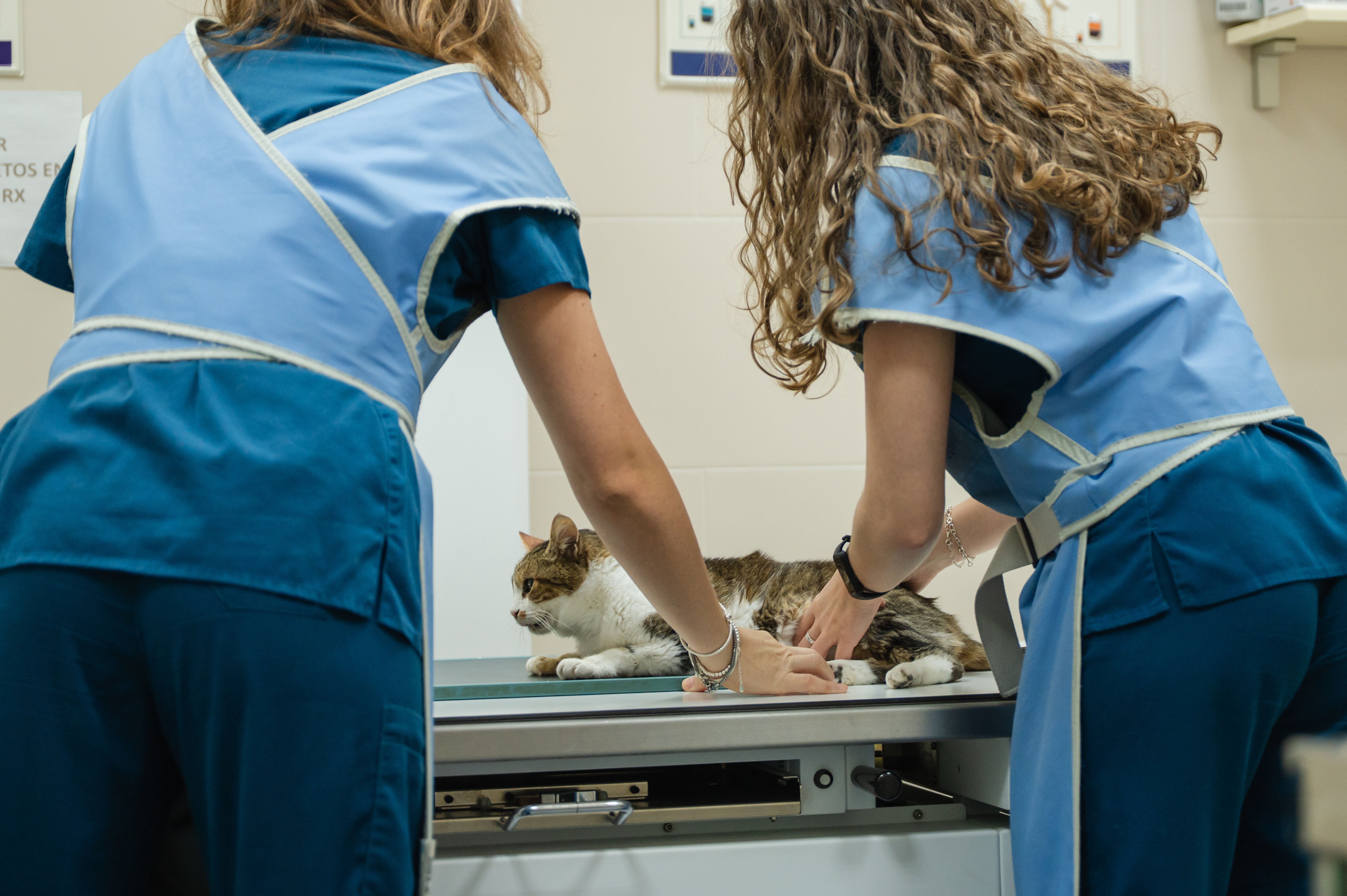
X-ray diagnostics are indispensable in routine veterinary practice. In practice, however, there is often a problem: Some animals cannot be sedated and must be held and restrained by the veterinarian, veterinary staff, or the pet owner during the imaging procedure. This leads to radiation exposure that is often underestimated. Three recent studies show that dosimetry, radiation protection measures, and increased awareness of actual radiation exposure are crucial for minimizing potential health risks.
To reliably determine radiation exposure, thermoluminescent dosimeters (TLDs) were used in the studies to quantify radiation doses to the extremities and organs. Measurements were taken both under and over protective clothing to assess the effectiveness of lead aprons, thyroid shields, and masks.
Radiation Exposure in Digital X-Ray Diagnostics
A recent study (2025)¹ determined radiation exposure during digital small-animal radiography in a total of 20 examinations. TLDs were placed on the animals’ skin, on the hands of the examiners, and under the lead aprons of the examiners. The mean entrance dose at the animal’s skin was 0.36 mGy. The average hand dose for the personnel holding the animal in place was measured at 0.35 mSv. The whole-body dose was 0.14 mSv. Radiation exposure to the hands was thus significantly higher, as the hands were at least partially directly exposed to the primary beam. The authors emphasized the need to consistently implement the ALARA principle (“As Low As Reasonably Achievable”) in veterinary practices. In addition to the consistent use of protective clothing, this also includes optimizing imaging protocols.¹
Scatter Radiation During Manual Animal Restraint
Oh et al. (2018)² investigated the radiation exposure of personnel restraining animals during 778 X-ray examinations of small animals. TLDs were attached to five anatomical sites (eyes, thyroid, chest, gonads, arm) both inside and outside the protective equipment. Without protection, the highest doses were measured at the eyes and thyroid; however, these could be reduced by 92% and up to 99%, respectively, through appropriate radiation protection. The hand dose could only be reduced by about 49% with appropriate protection – presumably due to the proximity to the primary beam. The study demonstrates that complete eye protection and the reduction of manual restraint are essential radiation protection measures.²
Fluoroscopic Examinations and Increased Hand Exposure
These findings are largely consistent with the study by An et al. (2019).3 In that study as well, the highest radiation dose was detected on the hands of the image-guidance technicians during 65 fluoroscopic examinations (up to 8.29 mSv per examination). The radiation dose to the eyes outside the shielding corresponded to approximately 25% of the recommended annual limit for the lens of the eye (20 mSv/year).4 The radiation protection equipment was most effective for the eyes (81.9% reduction), followed by the gonads (81%) and the chest (63.7%). In this study as well, the reduction at the hands was less pronounced (53.5%). Compared to other body parts, the radiation dose to the hands remained high even within the protective equipment, which can likely be attributed to scattered radiation penetrating through the open arm shields.3
Conclusion
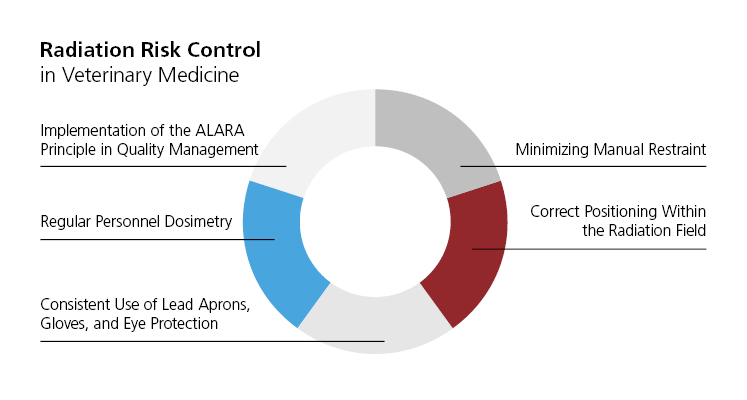
The risk of radiation exposure in veterinary imaging is real and primarily affects the eyes and hands. Although protective measures are extremely effective, they cannot completely prevent exposure. Therefore, according to the authors of the three studies, consistent protective strategies are necessary:
- Minimizing manual patient positioning
- Correct positioning within the radiation field
- Consistent use of eye protection, gloves, and lead aprons
- Regular dosimetry
- Implementation of the ALARA principle in in-house quality management
Sources
1Saeedian E, et al. Thermoluminescence dosimetry in small animal digital radiography, the dose to animals, and their adopters. Radioprotection 2025,60, 57-64.
2Oh H, et al. Restrainer exposure to scatter radiation in practical small animal radiography measured using thermoluminescent dosimeters. Veterinární medicína 2018,63, 81-6.
3An J, et al. Evaluation of radiation exposure from fluoroscopic examination in small animal veterinary staff using thermoluminescent dosimeters. Veterinární medicína 2019,64, 266-70.
4Stewart FA, et al. ICRP publication 118: ICRP statement on tissue reactions and early and late effects of radiation in normal tissues and organs--threshold doses for tissue reactions in a radiation protection context. Ann ICRP 2012,41, 1-322.
