How the radiologist arrives at his findings
Digital dictation
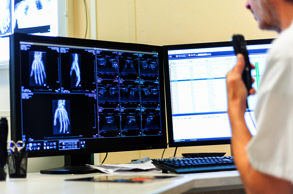
With digital dictation, the radiologist looks at the X-ray images and speaks his findings into special dictation devices or directly onto a smartphone or tablet. The voice files are then transmitted to typists via an encrypted and secure connection.
Compared to classic dictation, this procedure saves time because there is no need to transport cassettes and the digital images can be sent to transcriptionists and processed immediately. Radiologists can dictate at any time and any place. Digital dictation machines provide high-quality recordings, which facilitates transcription and reduces the susceptibility to errors. In addition, digital dictations can be seamlessly integrated into electronic health records (EHR) or other medical information systems such as RIS and PACS. This optimises the workflow and improves documentation.
Speech recognition
With speech recognition, the findings for imaging are dictated directly into a template of the hospital or radiology information system, corrected if necessary, then checked and released. The common speech recognition systems on the market can now handle dialects and the speech of foreign colleagues well.
It is obvious that the entire process of report preparation is thus significantly accelerated and radiologists can process more cases in less time in this way. The immediate control reduces the error rate considerably. If the software also offers the option of navigating in the menus by voice, radiologists can work even faster.
Structured Reporting
Structured reporting follows a fixed scheme with standardised templates for the preparation of X-ray reports. These templates contain predefined sections for clinical information, examination protocols, findings, diagnoses and conclusions, which the radiologist fills with text.
On the one hand, the standardised structure facilitates and accelerates the preparation of findings and, on the other hand, ensures a clear and uniform presentation of the findings, which improves readability and comprehensibility. Since important information on radiological diagnostics appears clearly, precisely and always in the same place in the report, this type of presentation improves communication between radiologists and other medical professionals. Although the structured reporting specifies where which information is recorded, it allows the radiologist to formulate it in his own words. Many doctors regard this freedom as an advantage.
Guided Reporting
However, this can also be a disadvantage, because it is difficult to evaluate an X-ray report with a large amount of free text. Guided Reporting therefore relies almost entirely on standardised entries via click boxes based on specific questions, defined answer options and checklists. Radiologists are guided step by step through the reporting process and it is ensured that all important information is systematically recorded.
The result is consistent and structured findings without free text in the essential sections. Findings created in this way are ideal for training artificial intelligence or for research purposes. As with structured reporting, automated analysis algorithms can be integrated, the results of which can be automatically transferred to the report. Overall, guided reporting offers the possibility of accelerating the entire process of establishing a diagnosis. For example, the structured templates, such as a normal finding, can be used as a starting point and specific information can be added or adapted. Due to the guided reporting, even less experienced radiologists are able to deliver reliable and high-quality results.